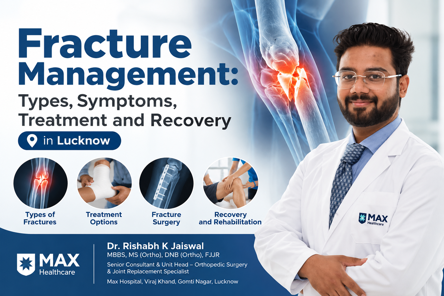
Introduction A fracture is a break or crack in a bone that can occur due to a fall, road traffic accident, sports injury or direct impact. Fractures can affect different parts of the body, including the arms, legs, wrists, ankles, hips and shoulders. Depending on the severity of the injury, a fracture may require a cast, splint, medication, rehabilitation or surgical treatment. Proper fracture management is essential to support bone healing, restore movement and reduce the risk of complications. Early diagnosis and appropriate treatment can help patients return to their daily activities safely. People searching for fracture treatment in Lucknow often want to understand the different types of fractures, available treatment options, the need for surgery and the expected recovery period. In this blog, we explain the causes, symptoms, diagnosis, treatment methods and rehabilitation process involved in fracture management. What Is Fracture Management? Fracture management refers to the medical assessment and treatment of a broken bone. The main objectives are to restore the bone’s alignment when necessary, maintain stability, support healing, manage pain and help the patient regain function. The appropriate treatment depends on several factors, including: Some fractures can heal with immobilisation using a cast or splint, while others require surgical procedures to stabilise the bone. An orthopaedic specialist evaluates these factors before recommending an appropriate treatment plan. What Are the Different Types of Fractures? Fractures are classified according to the pattern of the break, the extent of bone damage and whether the skin is affected. 1. Closed Fracture A closed fracture occurs when the bone breaks without creating an open wound through the skin. Although the skin remains intact, surrounding tissues may still be damaged. 2. Open Fracture An open fracture occurs when a wound communicates with the fracture, sometimes with the bone visible through the skin. This type of injury carries a significant risk of infection and requires emergency medical treatment. Do not attempt to push exposed bone back into the wound. Cover the wound with a clean dressing and seek emergency care. 3. Displaced Fracture A displaced fracture occurs when the broken bone fragments have moved out of their normal alignment. Depending on the injury, treatment may involve repositioning the bone or surgery. 4. Non-Displaced Fracture In a non-displaced fracture, the bone has broken but the fragments remain in relatively normal alignment. Many such fractures can be treated with appropriate immobilisation, depending on their location and stability. 5. Comminuted Fracture A comminuted fracture occurs when a bone breaks into three or more pieces. It can result from high-impact injuries and may require specialised treatment. 6. Stress Fracture A stress fracture is a small crack in a bone caused by repetitive loading or overuse. It commonly affects weight-bearing bones, particularly in people who increase their physical activity suddenly. 7. Greenstick Fracture A greenstick fracture is an incomplete fracture in which the bone bends and cracks rather than breaking completely. It is more common in children because their bones are more flexible. The type of fracture plays an important role in determining the treatment method and expected recovery. What Are the Common Causes of Bone Fractures? Bone fractures can occur because of sudden trauma or repeated stress on the bone. Common causes include: Identifying the cause helps the doctor assess the severity of the injury and determine whether additional investigations are needed. What Are the Symptoms of a Fracture? Fracture symptoms vary depending on the bone involved and the severity of the injury. Common signs include: 1. Pain A fracture often causes pain that worsens when the affected area is moved or placed under pressure. 2. Swelling Swelling may develop around the injured area shortly after the injury. 3. Bruising Blood vessels around the injured bone may be damaged, leading to bruising or discolouration. 4. Deformity The affected limb may look unusually shaped or appear out of alignment, particularly in displaced fractures. 5. Difficulty Moving or Bearing Weight A fracture in the leg, ankle or foot may make walking difficult. An arm fracture may interfere with lifting, gripping or other daily activities. 6. Tenderness The area around the fracture may be painful when touched. Not every fracture causes obvious deformity or severe pain. If a fracture is suspected following an injury, medical evaluation is important even when symptoms appear mild. When Should You Seek Emergency Fracture Treatment? Certain fractures require immediate medical attention to prevent serious complications. Seek emergency care if you notice: While waiting for medical help, keep the injured area as still as possible. Do not attempt to straighten the bone or push it back into place. If a serious injury is suspected, avoid unnecessary movement and follow emergency medical guidance. How Is a Fracture Diagnosed? Accurate diagnosis helps determine the appropriate treatment and reduces the risk of complications. 1. Medical History and Physical Examination The orthopaedic doctor will ask how the injury occurred, where the pain is located and whether the patient has any existing medical conditions. The examination may include checking swelling, tenderness, alignment, skin condition, circulation and nerve function. 2. X-ray An X-ray is commonly the first imaging test used to identify a fracture, assess bone alignment and determine the general pattern of the break. 3. CT Scan A CT scan may be recommended for complex fractures, particularly those involving joints or areas where detailed assessment of bone fragments is necessary. 4. MRI Scan An MRI may be useful when a stress fracture or an associated soft-tissue injury is suspected but is not clearly visible on an X-ray. 5. Additional Investigations Some patients may require blood tests or other investigations depending on the type of injury, planned surgery and general health. Not every patient requires all these tests. The doctor recommends investigations according to the clinical findings. What Are the Different Fracture Treatment Options? Fracture treatment depends on the bone involved, fracture stability, displacement, associated injuries and the patient’s health. 1. Cast or Splint Immobilisation A cast or splint keeps the injured bone stable while it heals. A cast may be recommended
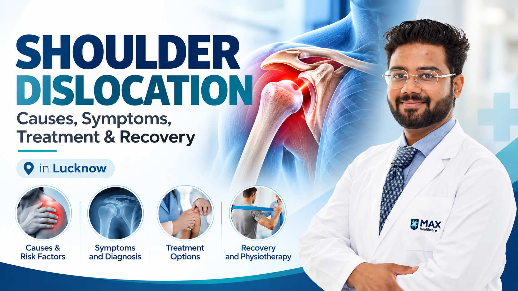
Shoulder Dislocation: Causes, Symptoms, Treatment and Recovery in Lucknow
Introduction Shoulder dislocation is a painful orthopaedic condition that occurs when the upper arm bone moves out of its normal position in the shoulder socket. It can happen following a fall, sports injury, road accident or sudden forceful movement of the arm. A dislocated shoulder can cause severe pain, swelling, visible changes in the shoulder’s shape and difficulty moving the arm. If you are experiencing shoulder pain or suspect a dislocation, timely medical assessment is important. Early diagnosis helps identify the extent of the injury and determine the appropriate treatment. People searching for shoulder dislocation treatment in Lucknow may have questions about the causes, symptoms, diagnosis, surgery and recovery process. Understanding these aspects can help patients make informed decisions about their orthopaedic care. At Max Hospital, Viraj Khand, Gomti Nagar, Lucknow, patients can seek an orthopaedic consultation to discuss their shoulder symptoms and appropriate treatment options with a qualified specialist. In this comprehensive guide, Dr. Rishabh K Jaiswal explains the important aspects of shoulder dislocation, from recognising the symptoms to understanding treatment and rehabilitation. What Is Shoulder Dislocation? The shoulder is a ball-and-socket joint that allows the arm to move in multiple directions. The rounded head of the upper arm bone, known as the humerus, fits into a shallow socket in the shoulder blade called the glenoid. The joint is stabilised by ligaments, muscles, tendons and a rim of cartilage called the labrum. These structures help maintain stability while allowing a wide range of movement. When the humeral head moves completely out of the socket, the condition is called a shoulder dislocation. If the joint moves only partially out of position, it is called a shoulder subluxation. Shoulder dislocation can affect people of different ages, including athletes, physically active individuals and older adults following a fall. The severity of the injury depends on the direction of the dislocation and whether the surrounding bones or soft tissues are damaged. If you experience a suspected dislocation, seek prompt medical attention rather than attempting to reposition the joint yourself. Types of Shoulder Dislocation Shoulder dislocation is classified according to the direction in which the upper arm bone moves. 1. Anterior Shoulder Dislocation Anterior shoulder dislocation is the most common type. In this condition, the humeral head moves forward out of the shoulder socket. It may occur when a person falls on an outstretched hand or experiences a forceful movement with the arm raised and rotated outward. Sports injuries and road accidents can also cause anterior shoulder dislocation. 2. Posterior Shoulder Dislocation Posterior shoulder dislocation occurs when the humeral head moves backward. It is less common and may be associated with seizures, electrical injuries or significant trauma. This type can sometimes be difficult to recognise without appropriate imaging. Prompt assessment is important to prevent delayed diagnosis. 3. Inferior Shoulder Dislocation Inferior shoulder dislocation is uncommon and occurs when the upper arm bone moves downward relative to the socket. It can result from substantial force applied to the arm and may be associated with injuries to surrounding nerves, blood vessels or other structures. The type of shoulder dislocation influences the investigations and treatment recommended by the orthopaedic specialist. What Are the Common Causes of Shoulder Dislocation? Shoulder dislocation can occur because of trauma, sports injuries or instability of the shoulder joint. Common causes include: Understanding the cause helps the doctor assess the injury and identify factors that may influence future shoulder stability. Symptoms of Shoulder Dislocation Recognising the symptoms of shoulder dislocation is important because the injury may require urgent medical attention. Common symptoms include: 1. Sudden and Severe Shoulder Pain A dislocated shoulder usually causes immediate pain. The discomfort may become worse when attempting to move the arm. 2. Visible Shoulder Deformity The shoulder may look different from the unaffected side. The normal rounded contour may appear flattened or altered. 3. Difficulty Moving the Arm Patients may find it difficult or impossible to raise, rotate or move the affected arm normally. 4. Swelling and Bruising Swelling and bruising can develop around the shoulder following the injury. 5. Numbness or Tingling Numbness, tingling or weakness in the arm or hand may indicate nerve involvement and requires urgent assessment. 6. A Feeling of Instability Some patients describe a sensation that the shoulder has slipped out of place or may dislocate again. If these symptoms develop after an injury, do not force the arm to move or attempt to push the joint back into position. Seek emergency medical care. When Should You Consult an Orthopaedic Doctor in Lucknow? A suspected shoulder dislocation should be assessed promptly by a qualified medical professional. Seek emergency care if you experience severe shoulder pain, visible deformity or inability to move the arm following an injury. A cold, pale or blue hand, reduced circulation, new numbness or significant weakness may indicate a serious complication. Patients who experience repeated shoulder dislocations, persistent instability or ongoing shoulder pain should also arrange an orthopaedic assessment. If you are looking for an orthopedic doctor in Lucknow for a shoulder injury, a clinical examination and appropriate imaging can help establish the diagnosis and guide treatment. Do not delay emergency care while trying to arrange a routine appointment. How Is Shoulder Dislocation Diagnosed? Diagnosis involves a medical history, physical examination and appropriate investigations. 1. Medical History and Physical Examination The doctor will ask how the injury occurred, whether the shoulder has dislocated previously and what symptoms are present. The examination may include checking the shoulder’s appearance, assessing safe movement where appropriate, and evaluating sensation, muscle function and blood circulation. 2. X-ray An X-ray is commonly used to confirm shoulder dislocation and identify associated fractures. Imaging also helps determine the direction of the dislocation. 3. MRI Scan Magnetic resonance imaging may be recommended when the doctor suspects damage to the labrum, ligaments, rotator cuff or other soft tissues. 4. CT Scan A CT scan may be required when there is suspected bone loss, a complex fracture or another injury that needs detailed assessment. Not every patient requires every